|
||
|
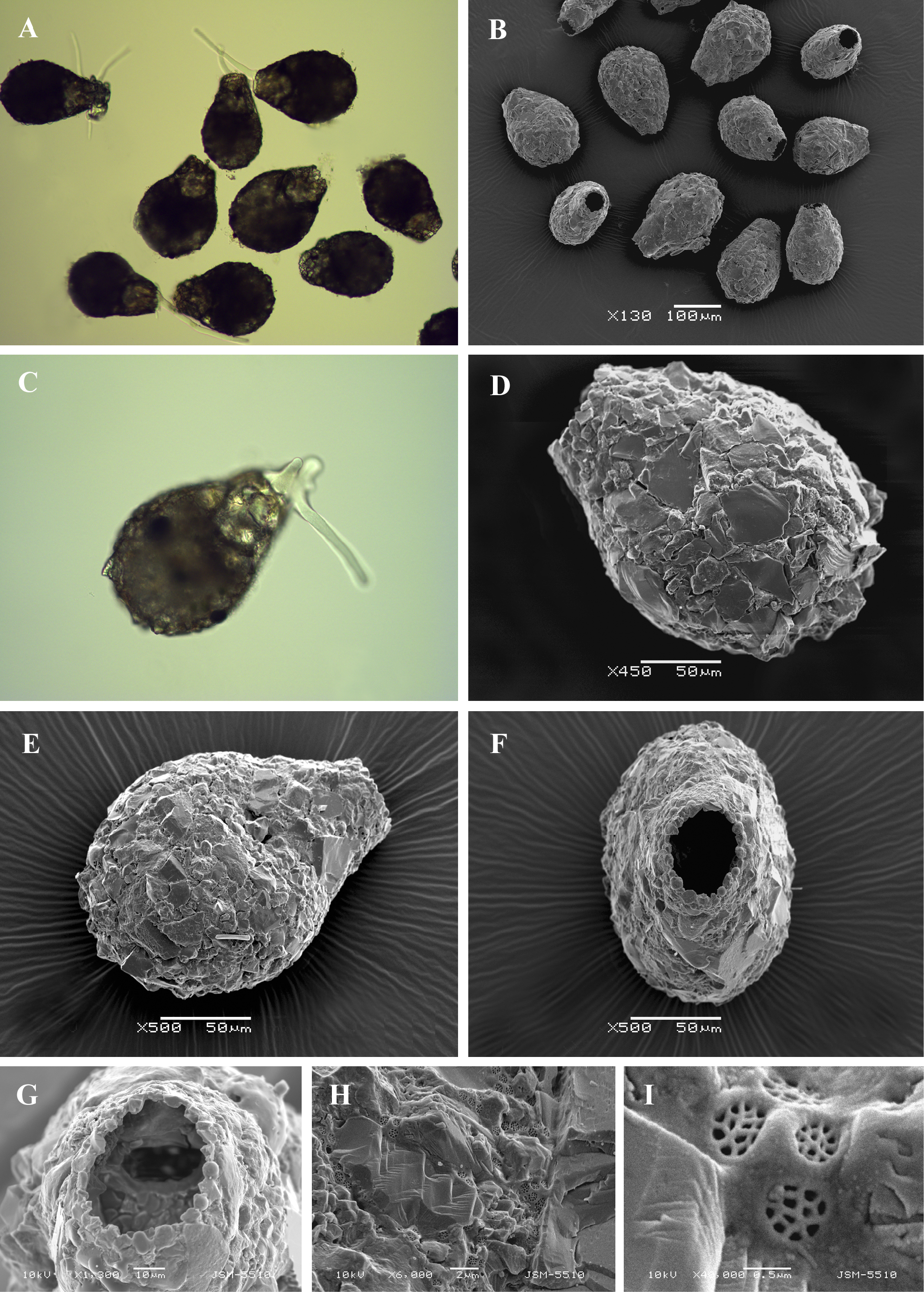
Light (A, C) and scanning electron (B, D-I) micrographs of Zivkovicia compressa. (A, B) View of many specimens to illustrate variability in shape and size of the shell. (C) View of live specimen showing endolobopodia. (D, E) Broad lateral views. (F) Apertural view (G) Apertural view of tilted specimen to show one of the internal openings. (H) Portion of shell showing its rough surface. (I) Detail of organic cement network. |