|
||
|
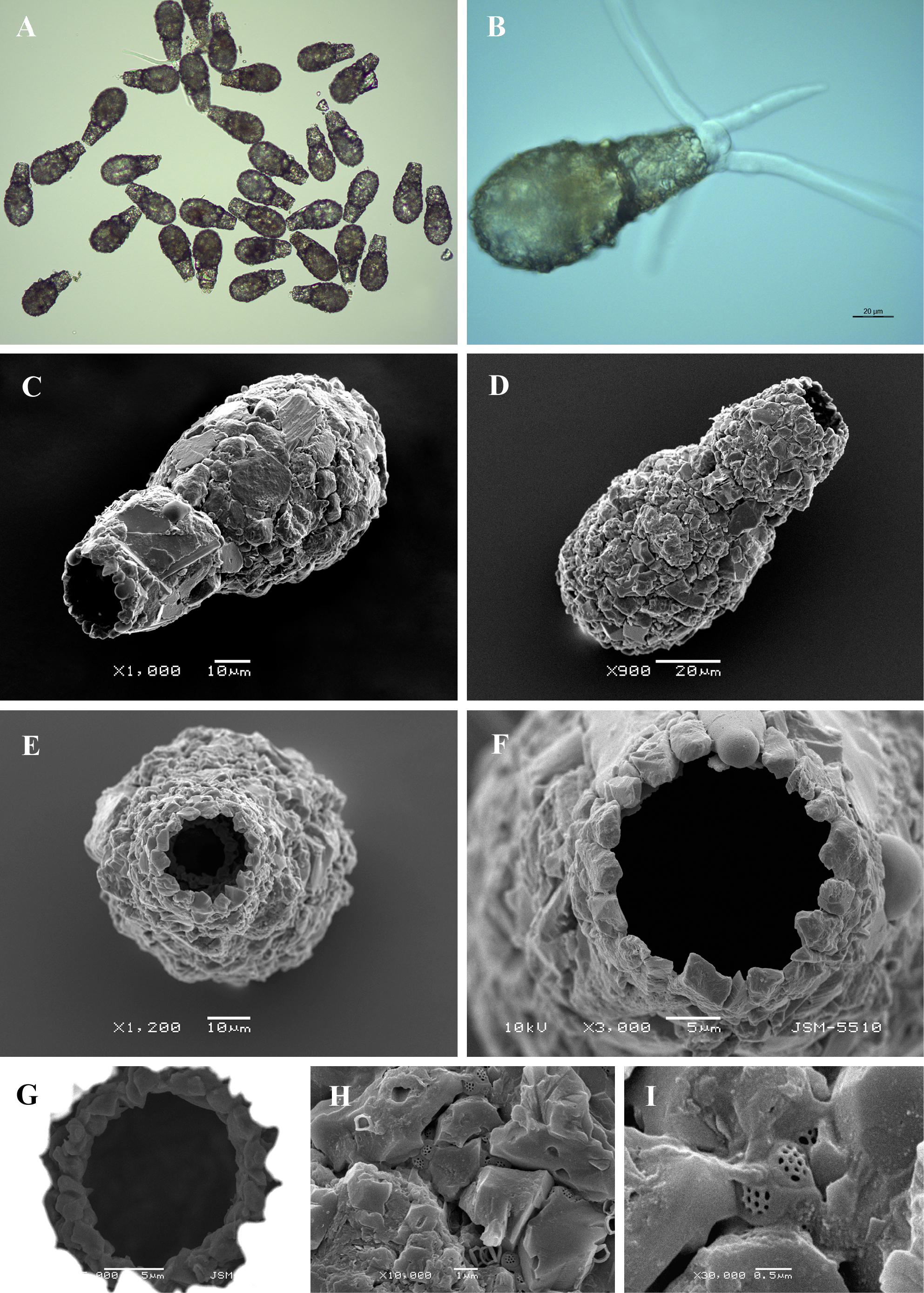
Light (A, B) and scanning electron (C-I) micrographs of Lagenodifflugia bryophila. (A) View of many specimens to illustrate variability in shape and size of the shell. (B) View of live specimen showing long endolobopodia and granular cytoplasm (C, D) Lateral view of two individuals to show a general shape. (E) Apertural view. (F) Close up view of aperture. (G) Close up view of internal opening. (H) Portion of shell to show arrangement of particles and rough surface of the shell. (I) Detail of organic cement network. |