|
||
|
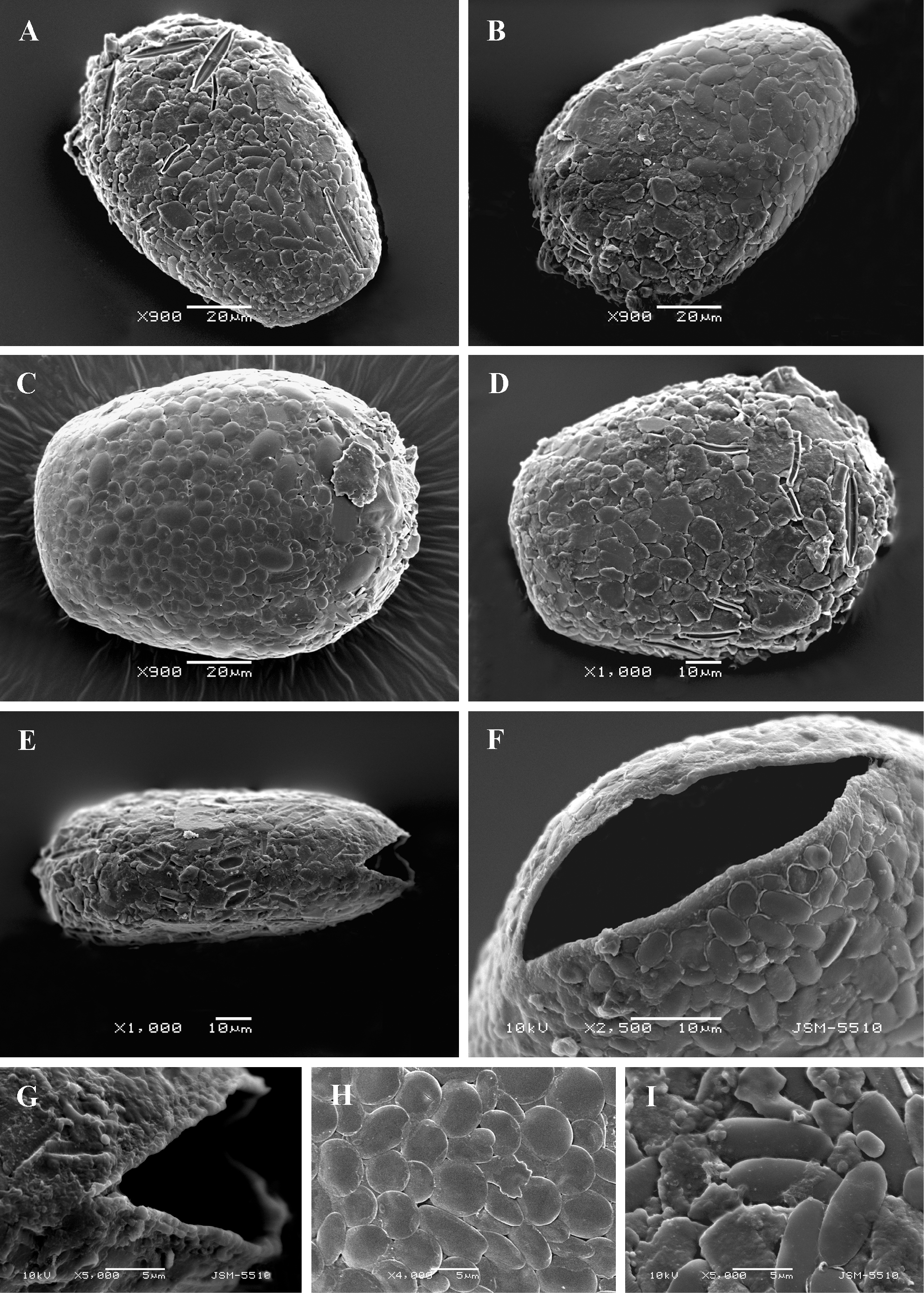
Scanning electron micrographs of Heleopera sphagni. (A-D) Broad lateral view of four specimens to illustrate variability in shape and structure of the shell. (E) Narrow lateral view. (F) Close up view of aperture to show a narrow elliptical aperture. (G) Lateral view showing acute commissures of aperture. (H, I) Details of shell surface showing differences in shape and arrangement of shell-plates. |