|
||
|
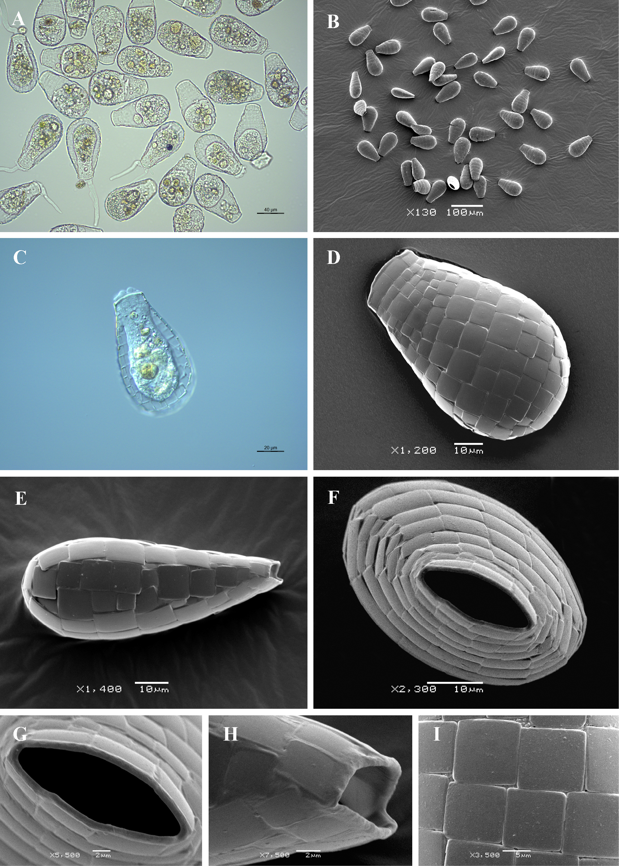
Light (A, C) and scanning electron (B, D-I) micrographs of Quadrulella symmetrica. (A, B) View of many specimens to illustrate variability in shape and size of the shell. (C) View of live specimen to show granular cytoplasm. (D) Broad lateral view. (E) Narrow lateral view. (F) Apertural view. (G) Close up view of aperture to illustrate its elliptical shape and bordering thin collar of organic cement (H) Lateral view of aperture. (I) Detail of shell surface showing shape and arrangement of shell-plates. |