|
||
|
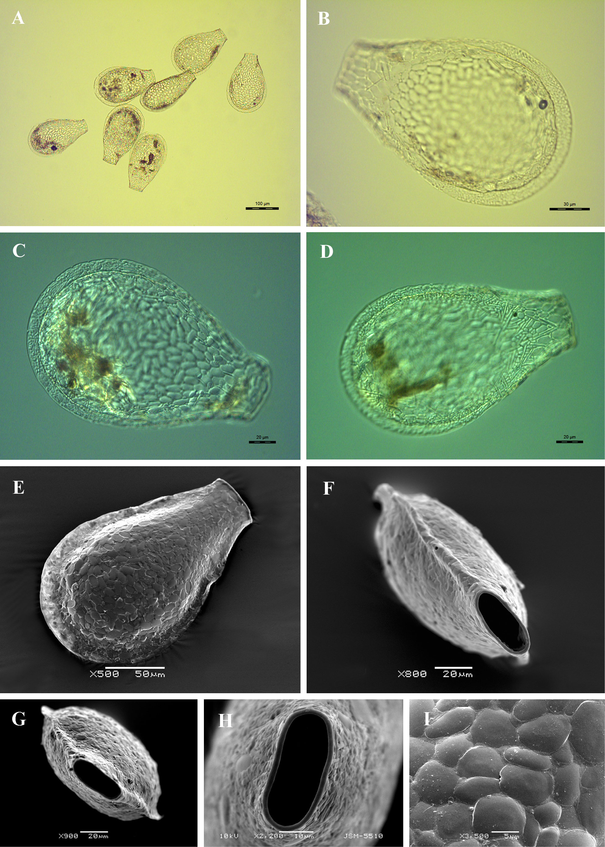
Light (A-D) and scanning electron (E-I) micrographs of Planocarina carinata. (A) View of several specimens to illustrate variability in shape and size of the shell. (B-E) Broad lateral view of four specimens showing general shape and distinct flattened lateral margin. (F) Latero-apertural view to illustrate flattened lateral margin and lateral pore. (G) Apertural view. (H) Close up view of aperture showing its oval outline and thin apertural collar. (I) Detail of shell surface to illustrate shape and arrangement of shell-plates in thick layer of organic cement. |