|
||
|
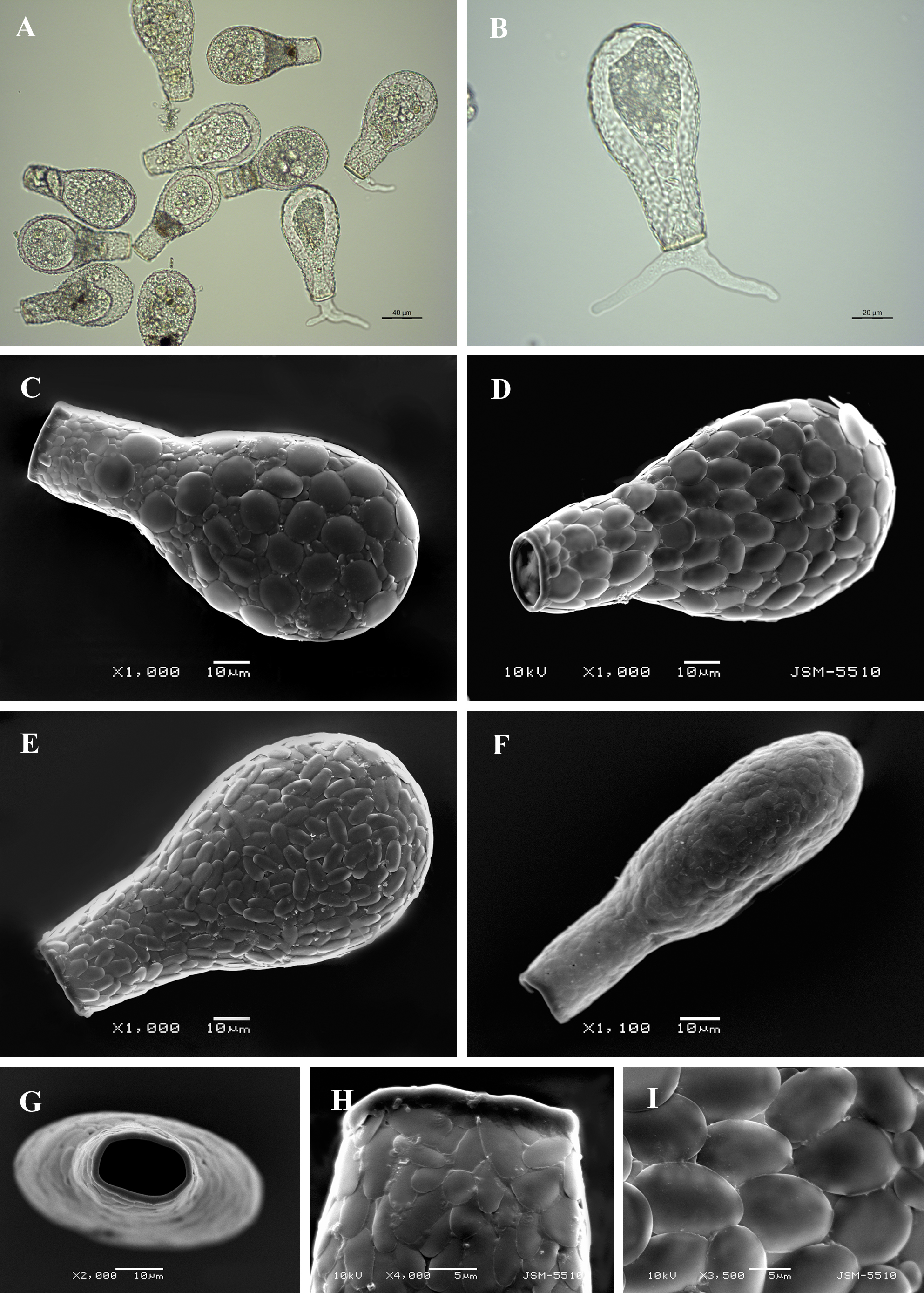
Light (A, B) and scanning electron (C-I) micrographs of Padaungiella wailesi. (A) View of several specimens to illustrate variability in shape and size of the shell. (B) View of live specimen showing granular cytoplasm and pseudopodia. (C-E) Broad lateral view of three specimens showing different shape and size of shell-plates. (F) Narrow lateral view. (G) Apertural view. (H) Broad lateral view of apertural region showing a thick collar of organic cement. (I) Detail of shell surface showing shape and arrangement of shell-plates. |