|
||
|
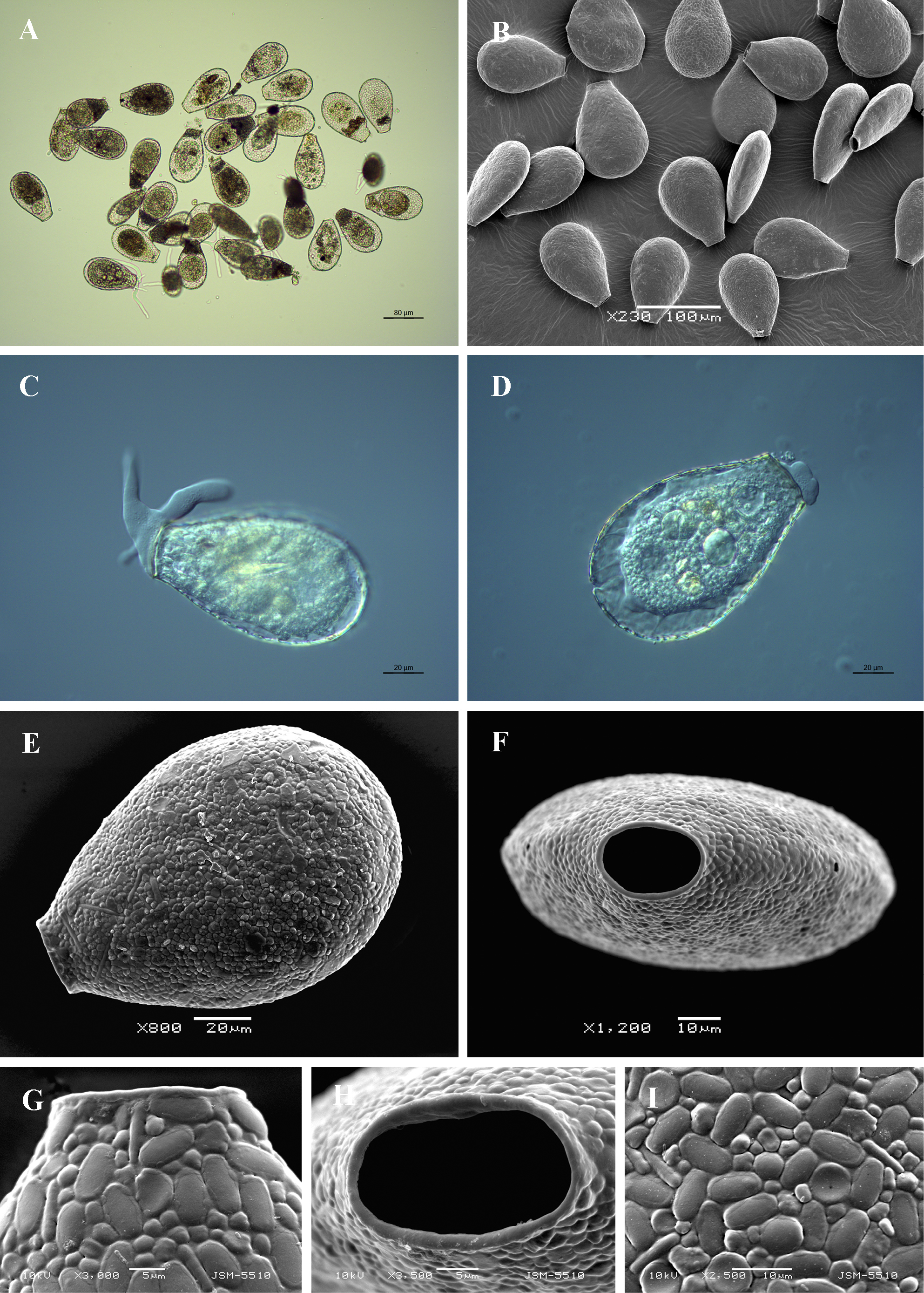
Light (A, C, D) and scanning electron (B, E-I) micrographs of Nebela collaris. (A, B) View of many specimens to illustrate variability in shape and size of the shell. (C, D) View of live specimens showing granular cytoplasm, pseudopodia and epipodes. (E) Broad lateral view. (F) Apertural view. (G) Broad lateral view of apertural region. (H) Close up view of aperture showing aperture outline and collar of organic cement. (I) Detail of shell surface showing shape and arrangement of shell-plates. |