|
||
|
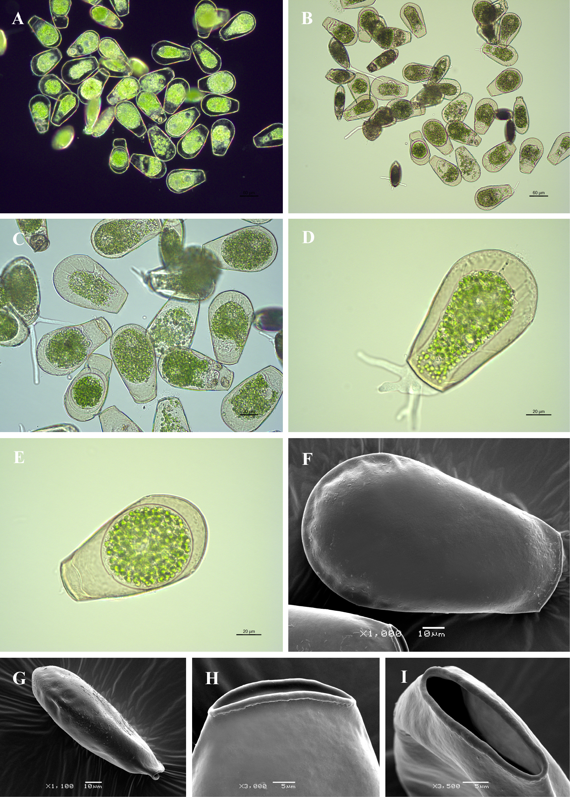
Light (A-E) and scanning electron (F-I) micrographs of Hyalosphenia papilio. (A-C) View of many specimens to illustrate variability in shape and size of the shell. (D) View of live specimen to illustrate the cytoplasm with symbiotic zoochlorellae, pseudopodia and epipodes. (E) View of encysted specimen showing round cyst. (F) Broad lateral view. (G) Narrow lateral view. (H) Broad lateral view of apertural region. (I) Close up view of aperture to illustrate its elliptical shape and bordering thick collar of organic cement. |