|
||
|
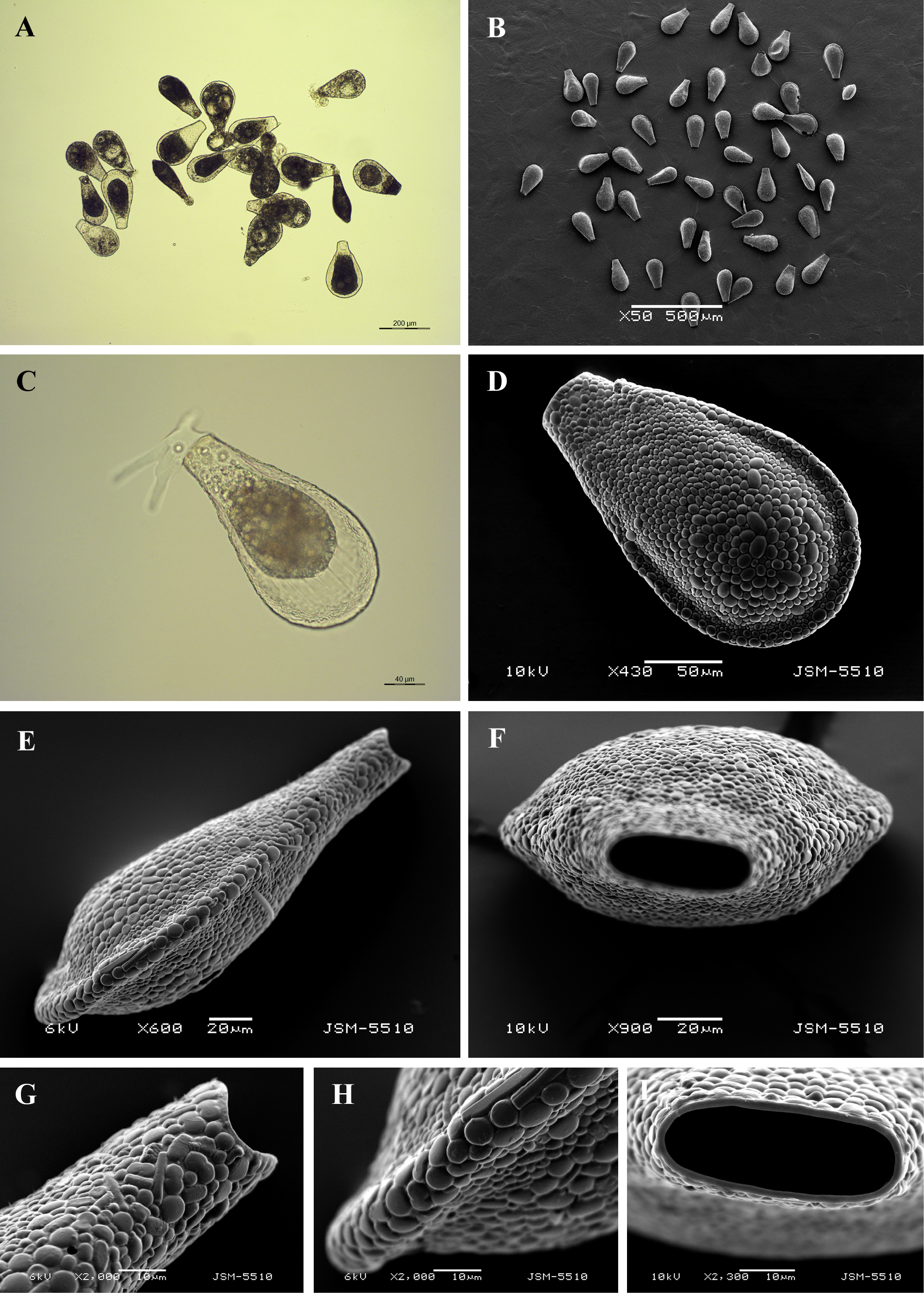
Light (A, C) and scanning electron (B, D-I) micrographs of Gibbocarina galeata. (A, B) View of many specimens to illustrate variability in shape and size of the shell. (C) View of live specimen to show granular cytoplasm, pseudopodia and epipodes. (D) Broad lateral view showing general shape. (E) Narrow lateral view to illustrate hollow tuberous keel. (F) Apertural view. (G) Lateral view of aperture. (H) Close up view of tuberous keel surrounding posterior end of the shell. (I) Close up view of aperture showing its oval outline and thin apertural collar. |