|
||
|
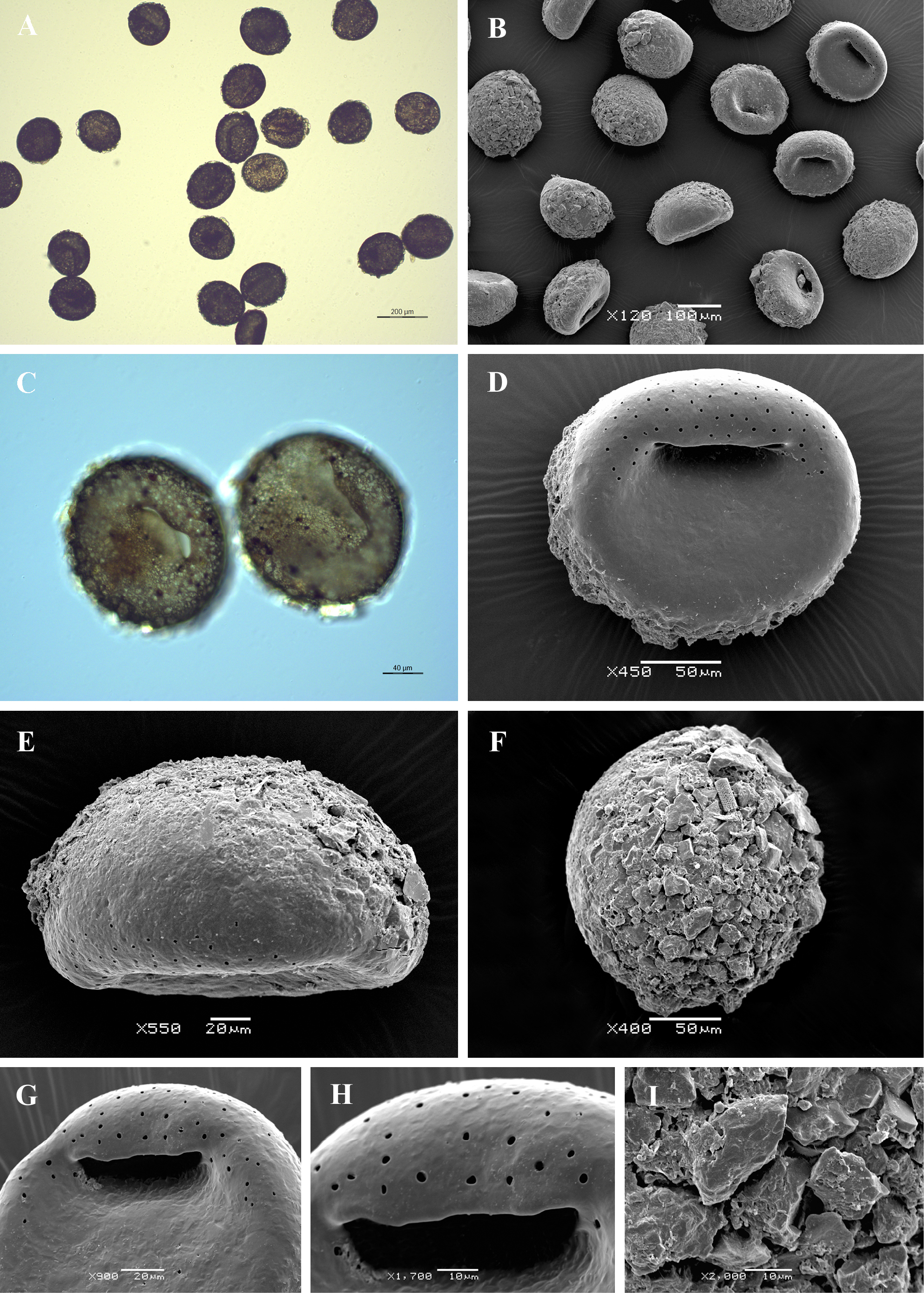
Light (A, C) and scanning electron (B, D-I) micrographs of Bullinularia indica. (A, B) View of many specimens to illustrate variability in shape and size of the shell. (C, D) Apertural views showing smooth apertural surface, characteristic aperture and large surrounding pores. (E) Lateral view. (F) Dorsal view. (G, H) Close up views of aperture to show its characteristic shape as an narrow, elongated slit. (I) Detail of dorsal side of the shell to illustrate its rough surface, covered with large pieces of quartz. |