|
||
|
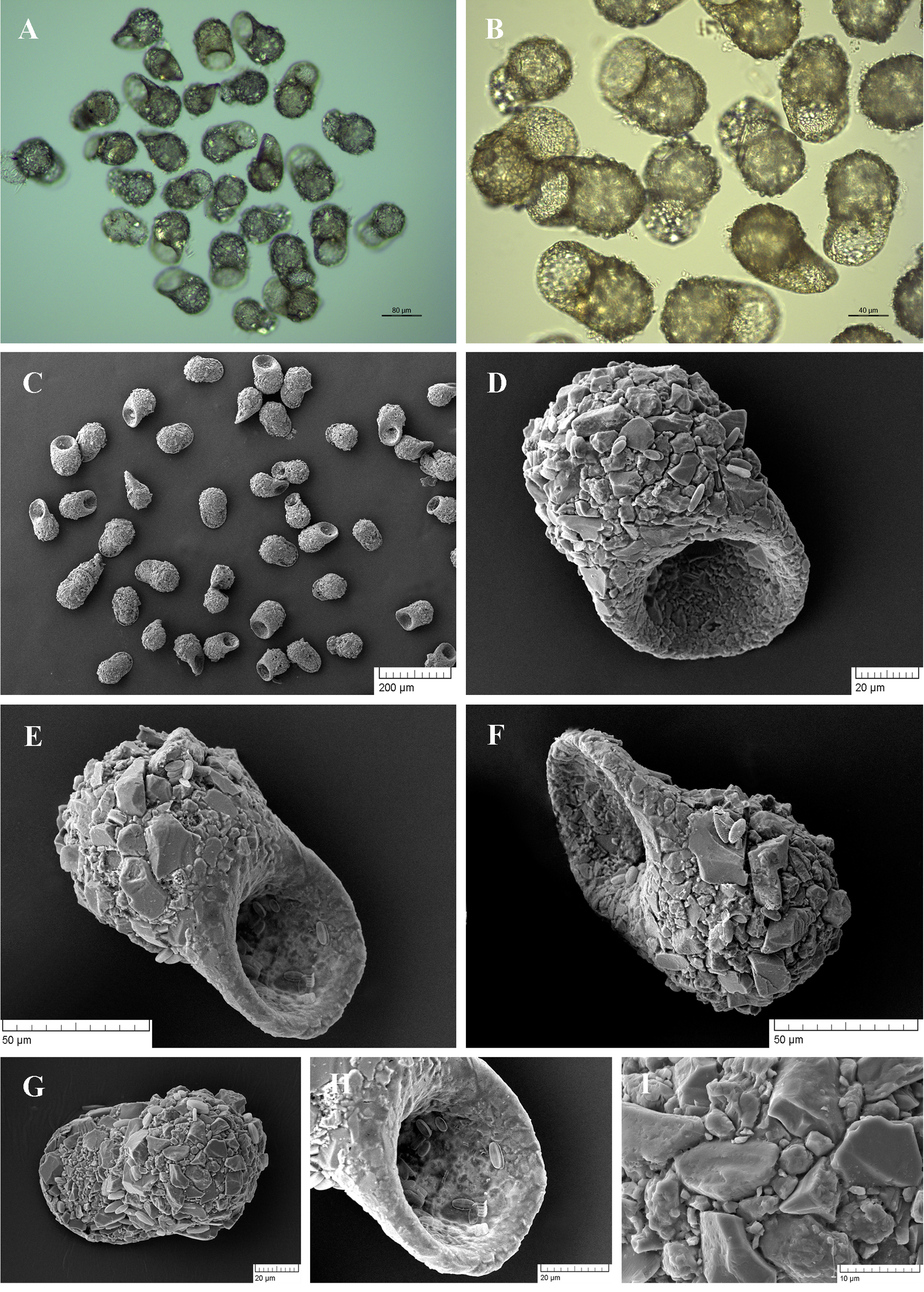
Light (A, B) and scanning electron (C-I) micrographs of Centropyxis platystoma. (A-C) View of many specimens to illustrate variability in shape and size of the shell. (D, E) Apertural view of two specimens showing general shape and shell structure. (F) Lateral view. (G) Dorsal view. (H) Close up view of aperture. (I) Detail of dorsal side of the shell to illustrate its rough surface, covered with medium to large pieces of quartz. |