|
||
|
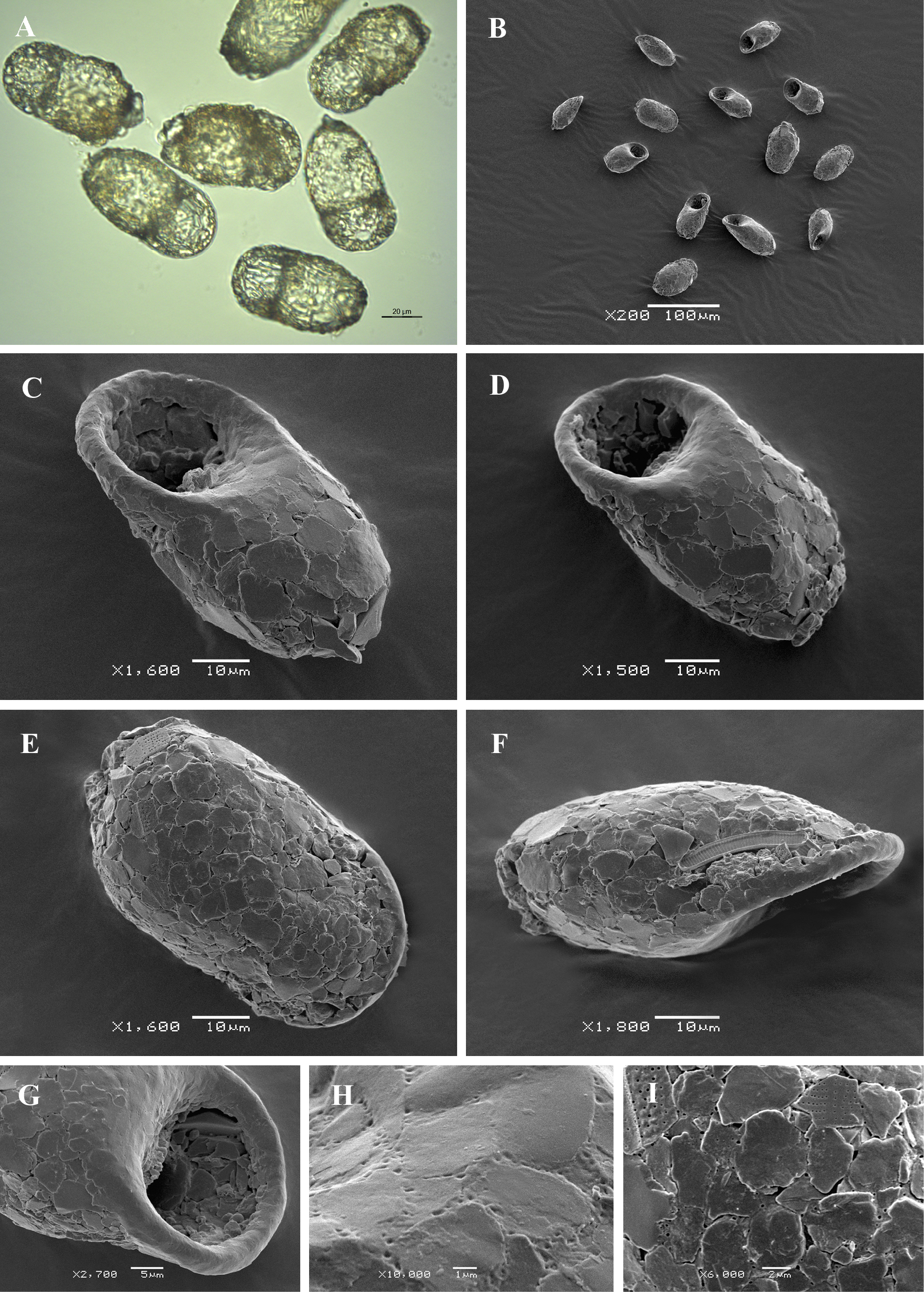
Light (A) and scanning electron (B-I) micrographs of Centropyxis elongata. (A, B) View of several specimens to illustrate variability in shape and size of the shell. (C, D) Apertural view of two specimens to show general shape and shell structure. (E) Dorsal view. (F) Lateral view. (G) Close up view of aperture. (H) Detail of smooth apertural surface. (I) Detail of aboral side of the shell to illustrate its rough surface, covered with flattish particles. |