|
||
|
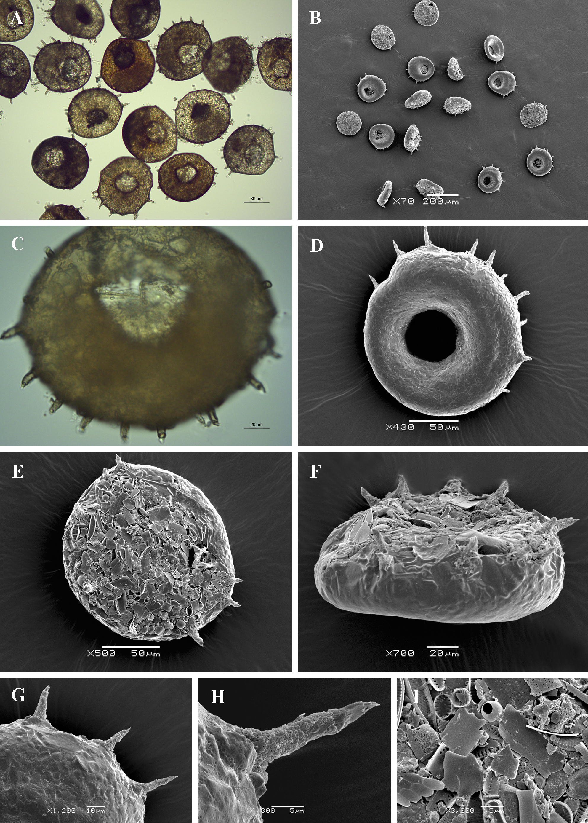
Light (A, C) and scanning electron (B, D-I) micrographs of Centropyxis discoides. (A, B) View of many specimens to illustrate variability in shape and size of the shell. (C, D) Apertural view of two specimens to illustrate general shape and shell structure. (E) Dorsal view. (F) Lateral view. (G) Close up view showing smooth apertural surface and disposition of spines on the lateral margin. (H) Close up view of a single spine to show its structure. (I) Detail of dorsal side of the shell to illustrate its rough surface, covered with sand grains and diatom frustules. |