|
||
|
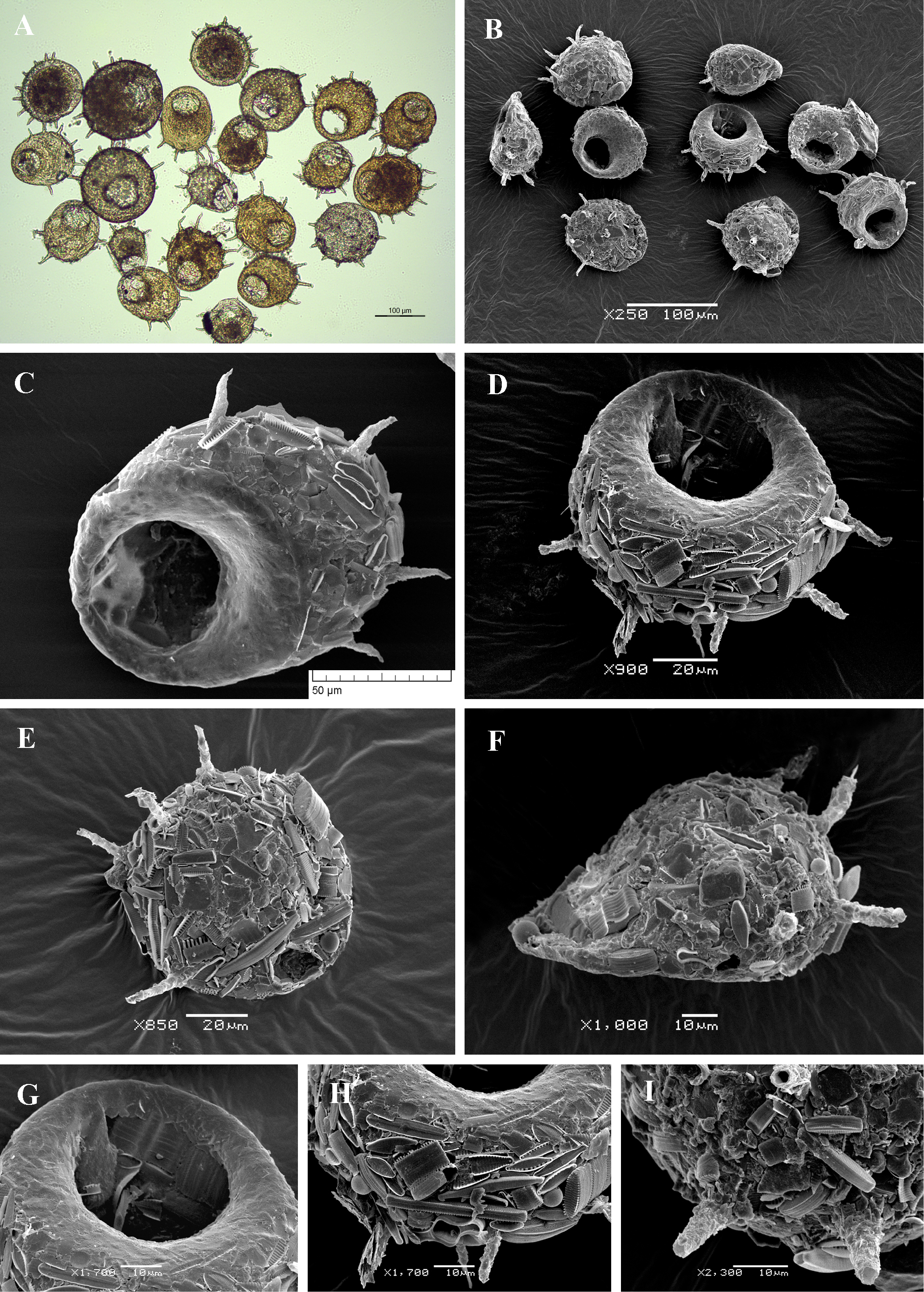
Light (A) and scanning electron (B-I) micrographs of Centropyxis aculeata. (A, B) View of many specimens to illustrate variability in shape and size of the shell. (C, D) Apertural view of two specimens to illustrate general shape and shell structure. (E) Aboral view. (F) Lateral view. (G) Close up view of aperture to illustrate its circular shape and smooth surface around the aperture. (H, I) Details of posterior end of the shell to illustrate its rough surface, covered with sand grains and diatom frustules. |