|
||
|
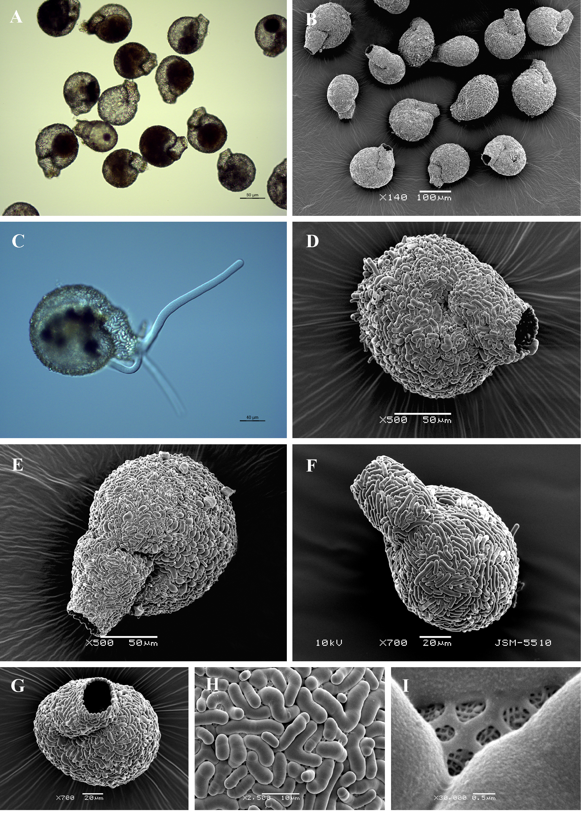
Light (A, C) and scanning electron (B, D-I) micrographs of Lesquereusia gibbosa. (A, B) View of many specimens to illustrate variability in shape and size of the shell. (C) View of live specimen showing granular cytoplasm and long pseudopodia. (D, E) Broad lateral view of two individuals to show general shape. (F) Narrow lateral view. (G) Apertural view. (H) Detail of shell surface showing shape and arrangement of siliceous curved rods and network of organic cement. (I) Close up view of a network of organic cement. |