|
||
|
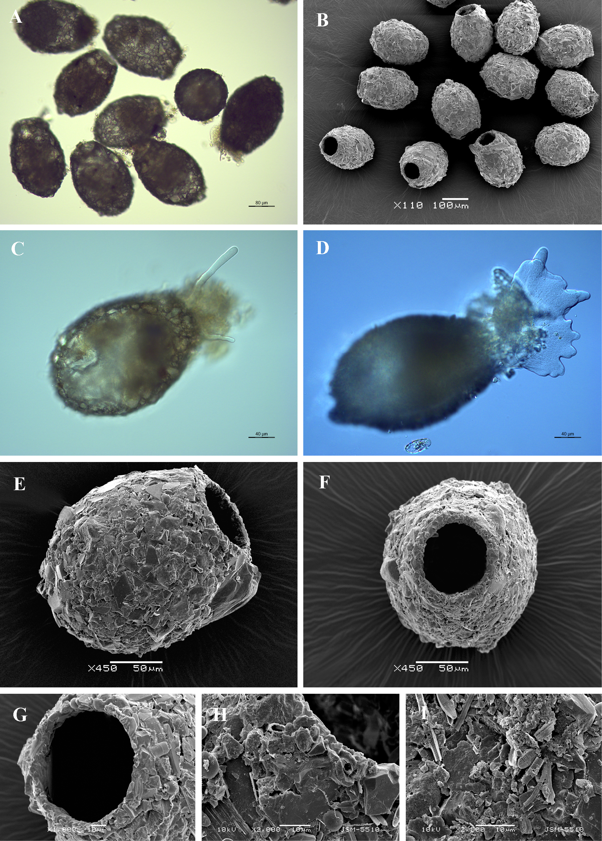
Light (A, C, D) and scanning electron (B, E-I) micrographs of Difflugia viscidula. (A, B) View of several specimens to illustrate variability in shape and size of the shell. (C-D) Lateral view of two live specimens showing different types of pseudopodia. (E) Lateral view. (F) Apertural view. (G) Close up view of aperture to show its regular outline. (H) Lateral view of apertural region. (I) Portion of shell to show arrangement of particles and rough surface of the shell. |