|
||
|
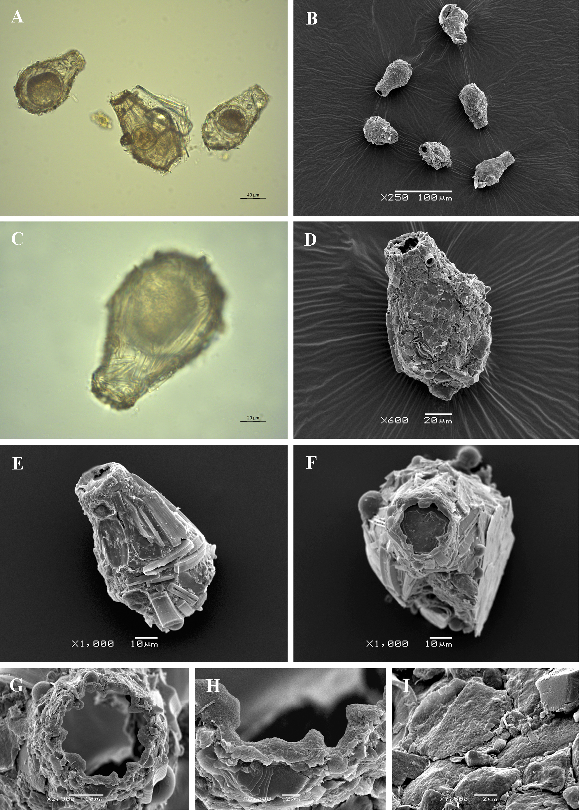
Light (A, C) and scanning electron (B, D-I) micrographs of Difflugia rubescens. (A, B) View of several specimens to illustrate variability in shape and size of the shell. (C) View of encysted specimen. (D-E) Lateral view of two specimens to illustrate rough surface of the shell. (F) Apertural view. (G-H) Close up view of aperture showing the bordering crenulated organic collar. (I) Portion of shell to show arrangement of particles and rough surface of the shell. |