|
||
|
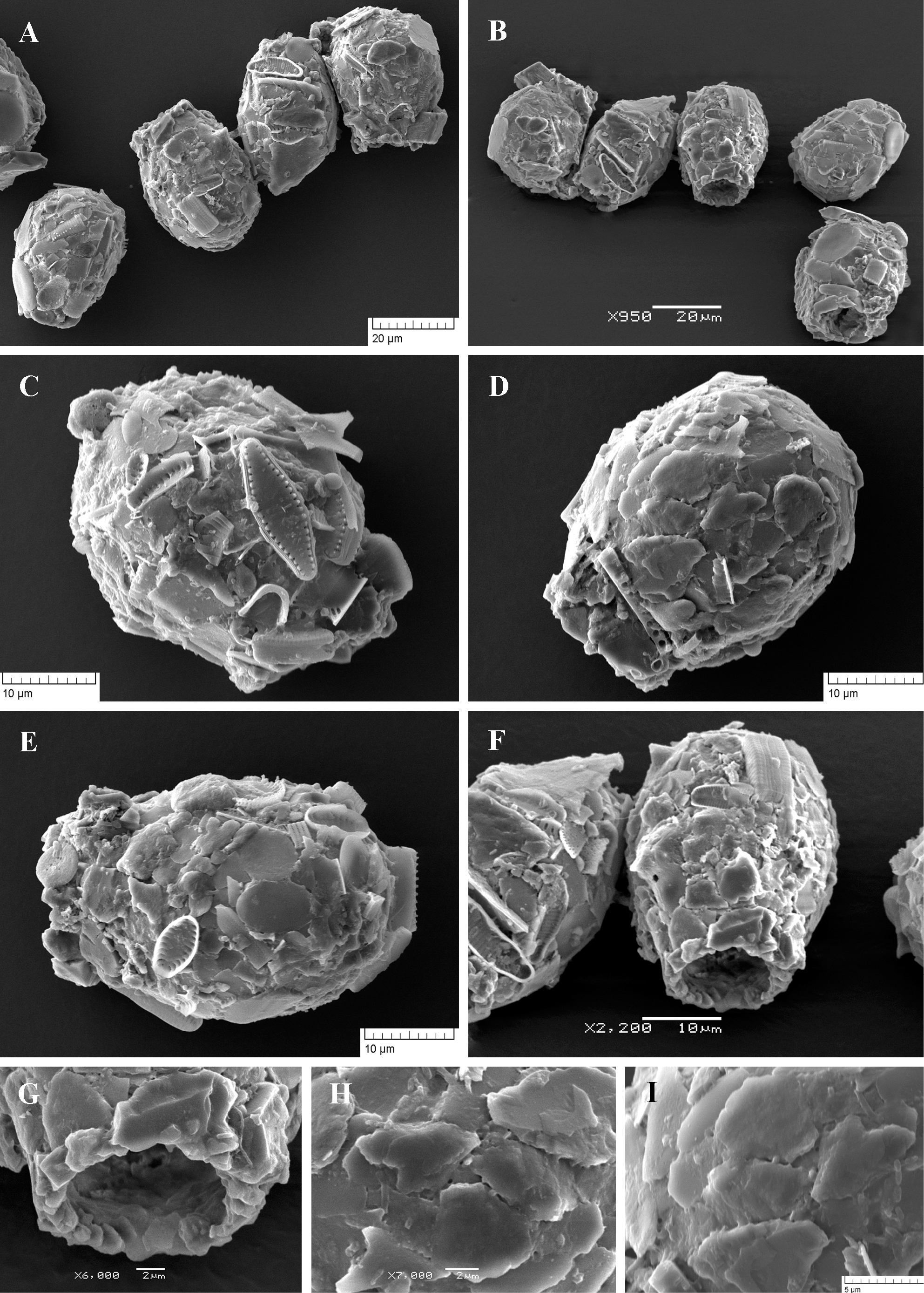
Scanning electron micrographs of Difflugia pulex. (A, B) View of several specimens to illustrate variability in shape and size of the shell. (C-E) Lateral view of three specimens showing different shape and shell structure. (F) Latero-apertural view. (G) Apertural view to show its irregular edge. (H, I) Portion of shell surface to show arrangement of flattish particles and small areas of organic cement with single small pores. |