|
||
|
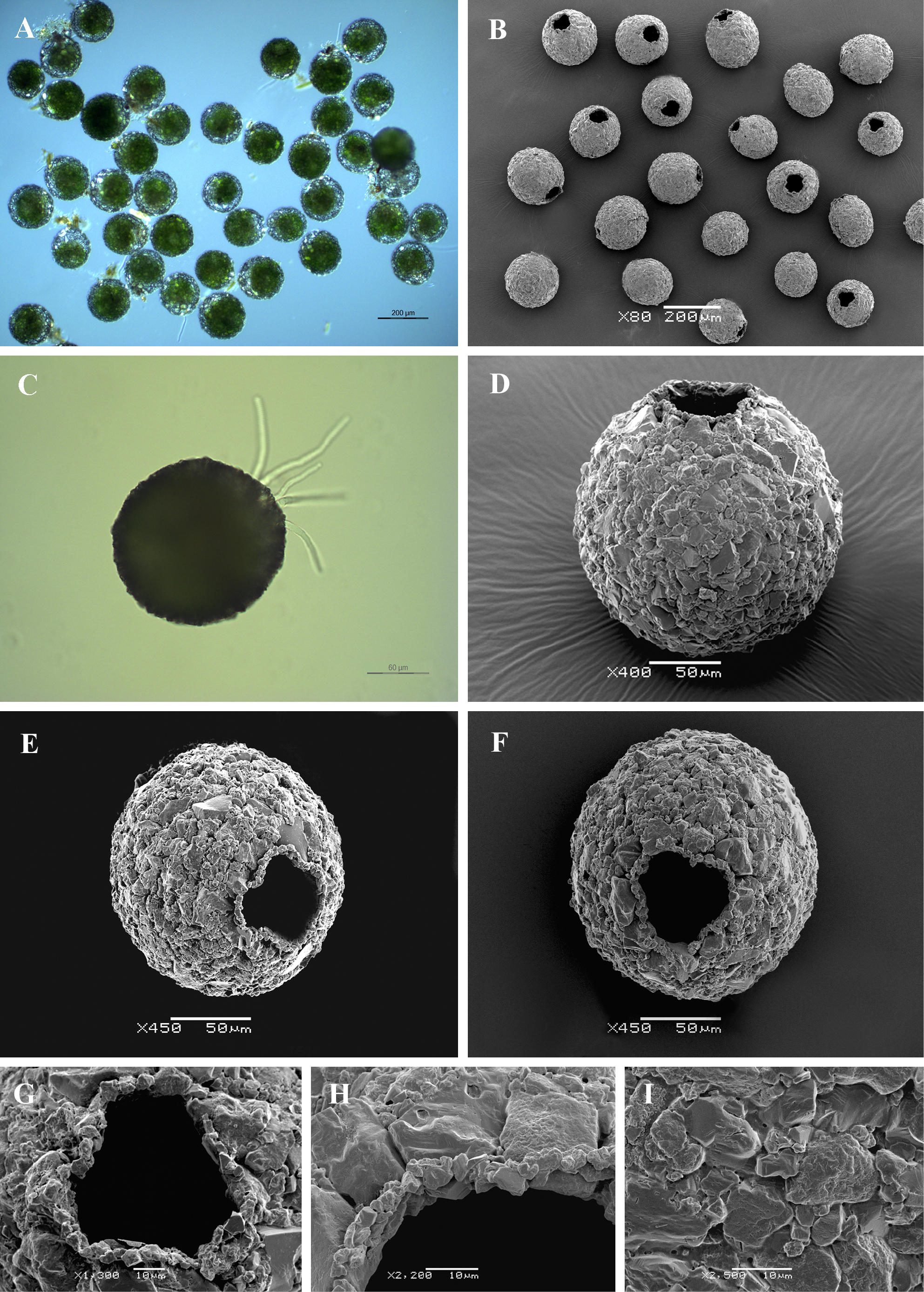
Light (A, C) and scanning electron (B, D-I) micrographs of Difflugia lobostoma. (A, B) View of many specimens to illustrate variability in shape and size of the shell. (C) View of live specimen to illustrate numerous pseudopodia. (D) Lateral view. (E, F) Apertural view of two specimens showing general shape, as well as tri- and four lobed aperture. (G, H) Close up views of aperture to illustrate its rough rim and trilobed aperture. (I) Portion of shell to illustrate its rough surface, covered with sand grains. |