|
||
|
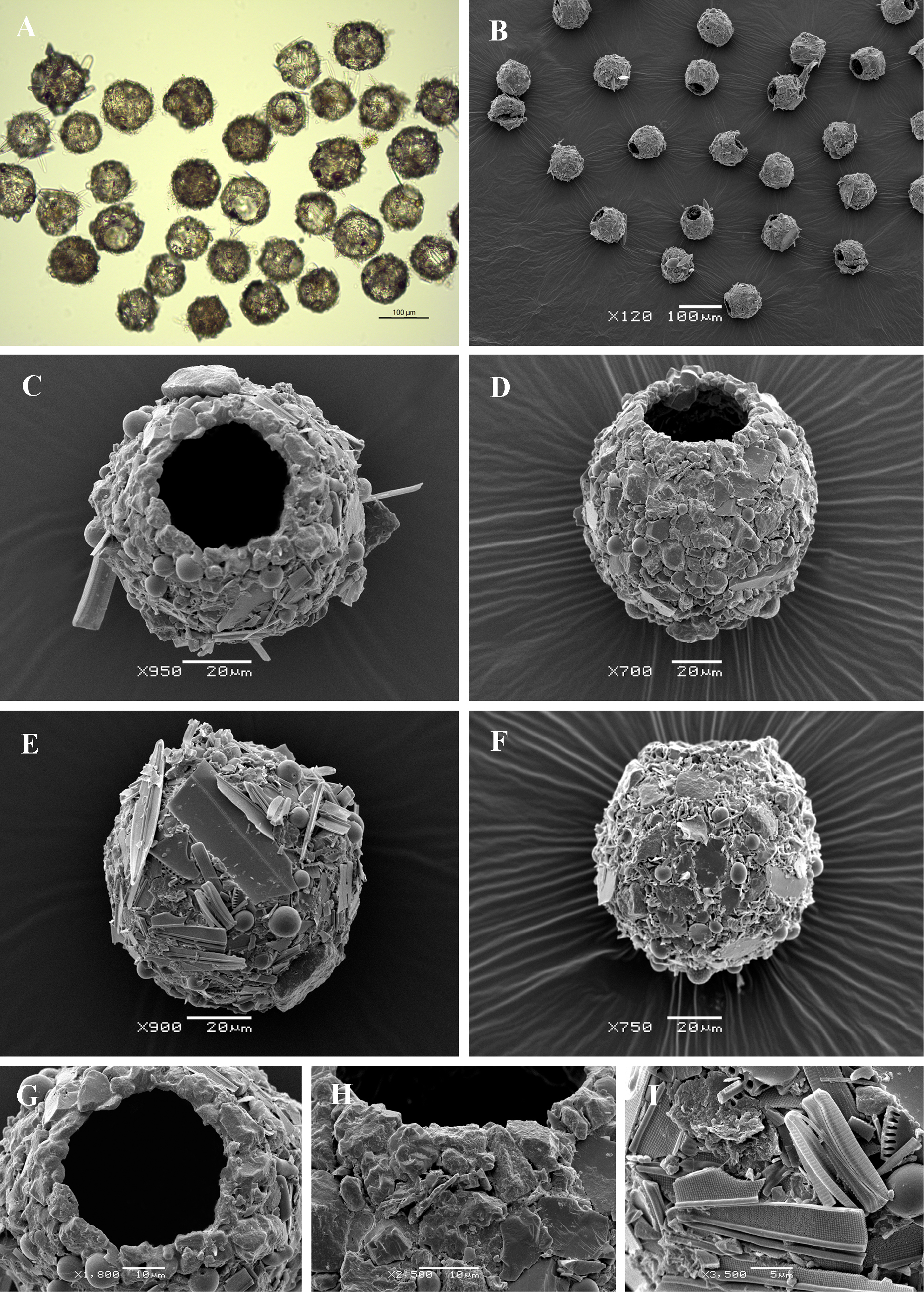
Light (A) and scanning electron (B-I) micrographs of Difflugia globulosa. (A, B) View of many specimens to illustrate variability in shape and size of the shell. (C-D) Apertural view of two specimens showing general shape. (E) Dorsal view. (F) Lateral view. (G, H) Close up views of aperture to illustrate its rough rim and circular outline. (I) Detail of aboral side of the shell to illustrate its rough surface, covered with sand grains and diatom frustules. |