|
||
|
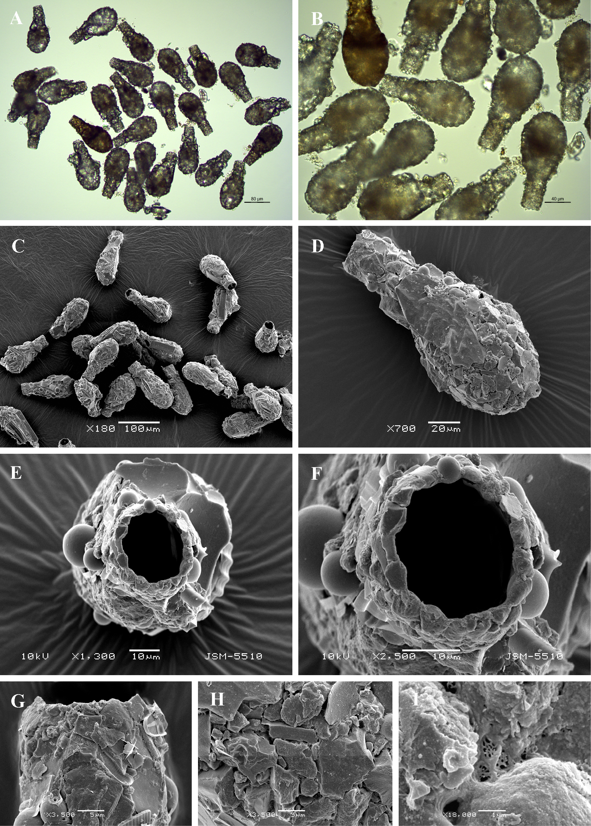
Light (A, B) and scanning electron (C-I) micrographs of Difflugia bryophila. (A-C) View of many specimens to illustrate variability in shape and size of the shell. (D) Lateral view showing rough surface of the shell. (E) Apertural view. (F) Close up view of aperture to show bordering particles. (G) Lateral view of apertural rim showing its irregularity (H) Portion of shell to show arrangement of particles and rough surface of the shell. (I) Detail of organic cement network. |