|
||
|
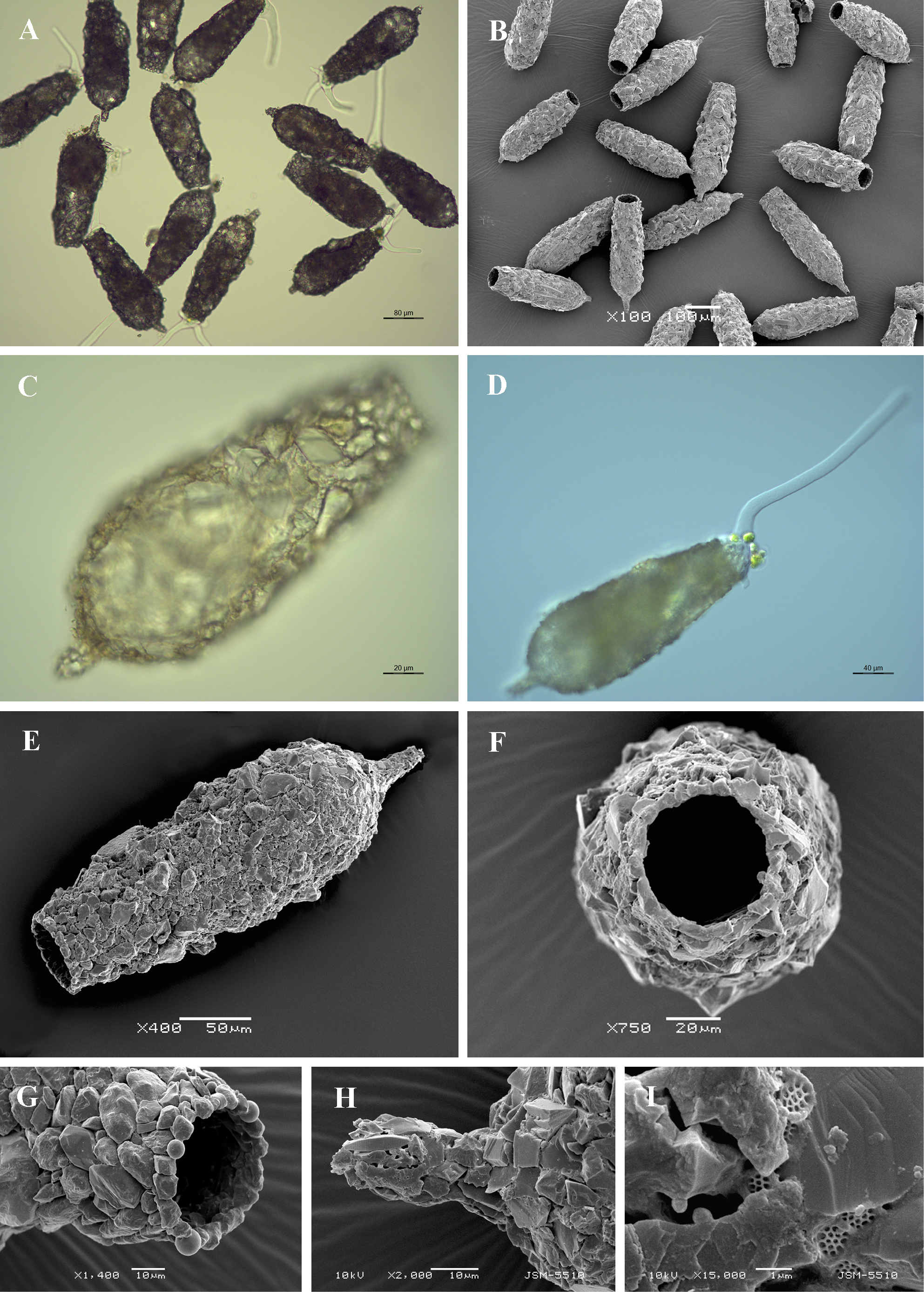
Light (A, C, D) and scanning electron (B, E-I) micrographs of Difflugia acuminata. (A, B) View of many specimens to illustrate variability in shape and size of the shell. (C, E) Lateral views of two specimens showing general shell shape. (D) View of live specimen to illustrate a single long endolobopodia. (F) Apertural view showing regular apertural outline. (G) Latero-apertural view to show apertural rim and surrounding, regularly arranged small particles (H) View of acuminate aboral region. (I) Detail of organic cement network. |