|
||
|
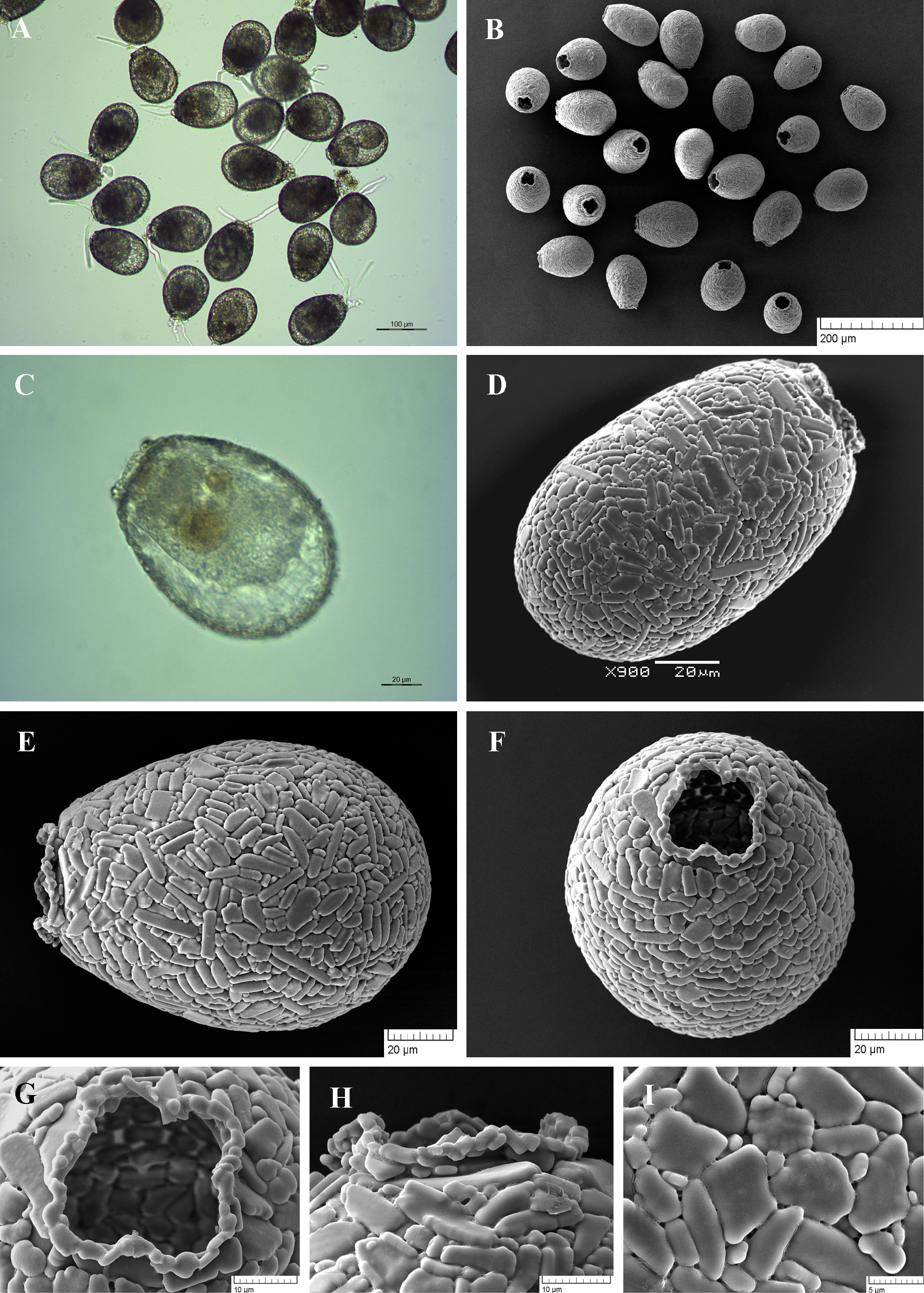
Light (A, C) and scanning electron (B, D-I) micrographs of Netzelia oviformis. (A, B) View of many specimens to illustrate variability in shape and size of the shell. (C) View of live specimen to show numerous epipodes. (D, E) Lateral views of two specimens to show general shape. (F) Apertural view. (G) Close up view of aperture to illustrate four lobes of aperture and narrow collar of small siliceous particles. (H) Close up view of a narrow collar of small pieces of quartz. (H) Detail of shell surface to illustrate regular arrangement of siliceous particles. |