|
||
|
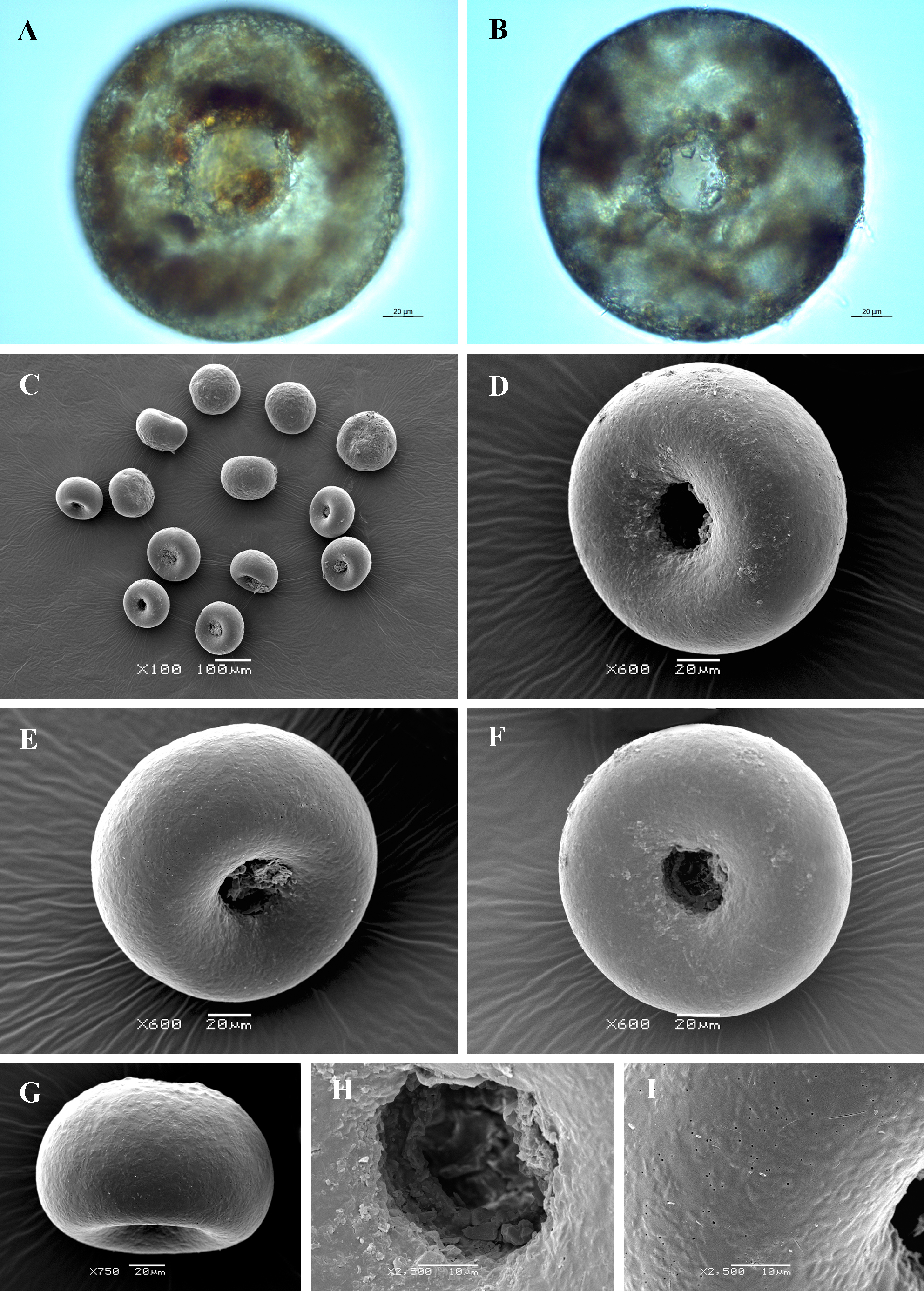
Light (A, B) and scanning electron (C-I) micrographs of Cyclopyxis puteus. (A, B) Light micrographs showing general shape. (C) View of many specimens to illustrate variability in shape and size of the shell (D-F) Apertural view of three specimens showing smooth apertural surface and deeply invaginated aperture. (G) Lateral view. (H) Close up view of aperture to illustrate its apertural tube and internal opening. (I) Detail of apertural side to illustrate its smooth surface and numerous pores in the organic cement. |