|
||
|
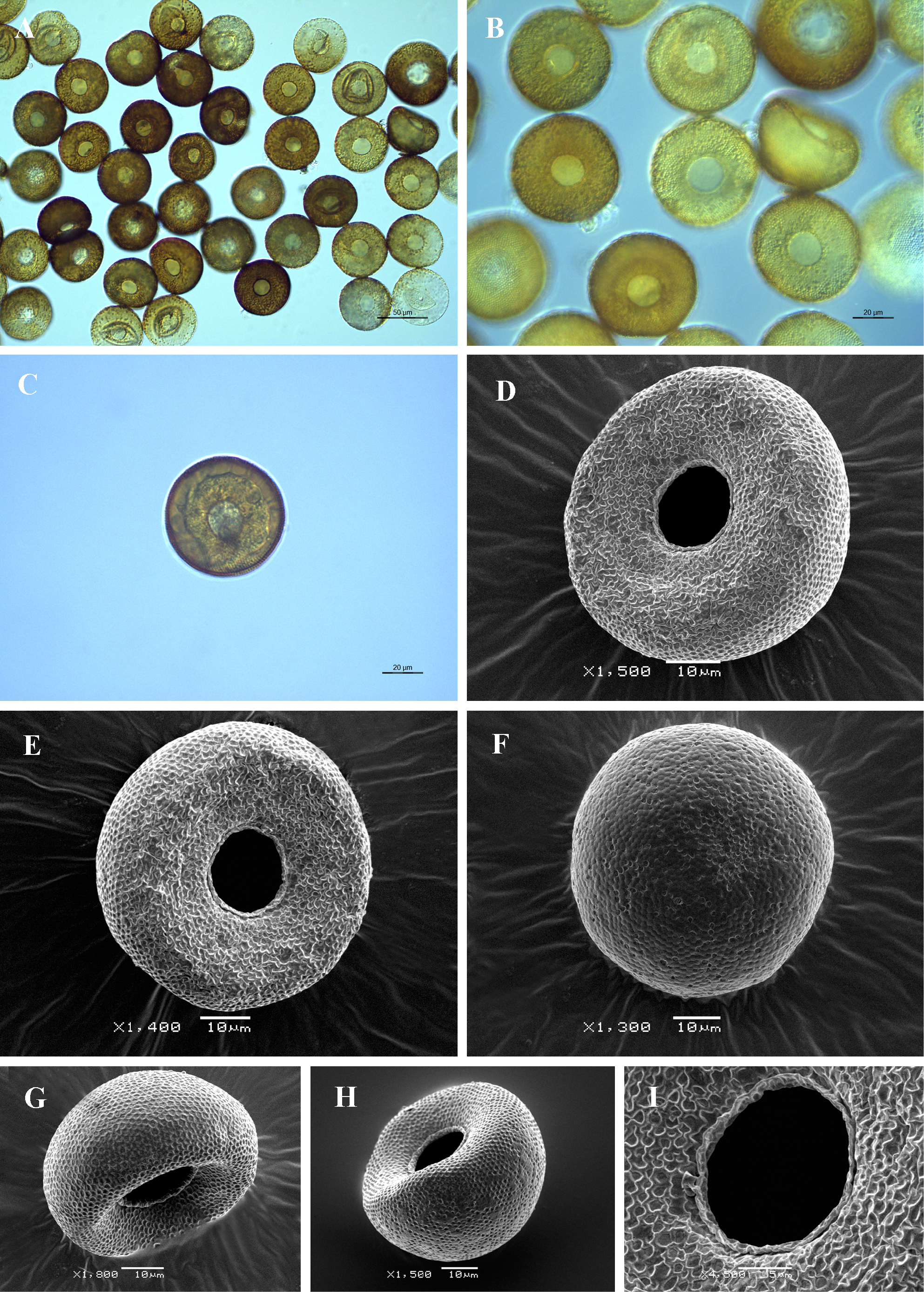
Light (A-C) and scanning electron (D-I) micrographs of Arcella hemisphaerica. (A, B) View of many specimens showing variability in shape and size of the shell. (C) View of live specimen illustrating numerous short epipodes and two typical vesicular nuclei with one large central nucleolus. (D, E) Apertural view of two specimens. (F) Dorsal view. (G, H) Latero-apertural view. (I) Close up view of aperture showing the apertural lip, irregular shell surface caused by drying of the shell and collapsed alveoli, as well as numerous small pores. |