|
||
|
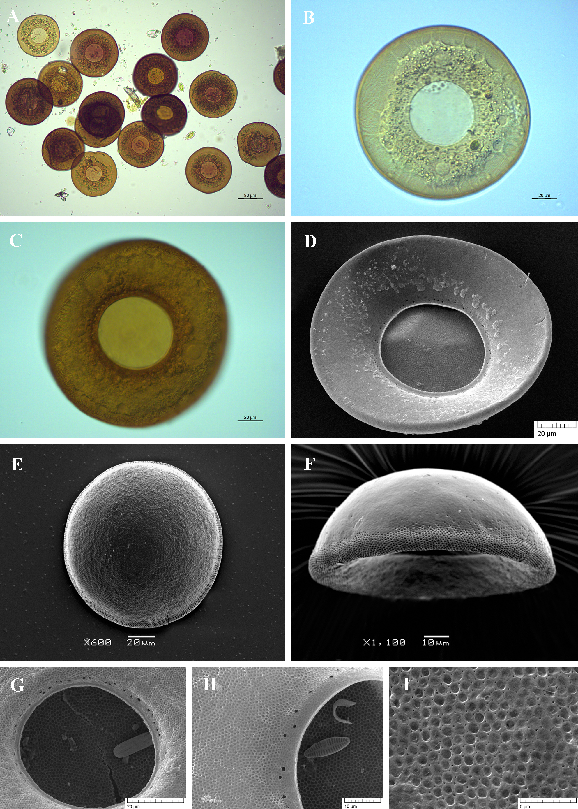
Light (A-C) and scanning electron (D-I) micrographs of Arcella discoides. (A) View of many specimens showing variability in shape and size of the shell. (B, C) View of live specimens illustrating numerous short epipodes and two typical vesicular nuclei with one large central nucleolus. (D) Apertural view to show smooth apertural surface, large aperture and surrounding numerous pores. (E) Aboral view. (F) Lateral view. (G, H) Close up view of aperture to show its circular outline, thin apertural lip and numerous surrounding large pores. (I) Portion of shell surface showing small organic alveoli on the aboral surface. |