|
||
|
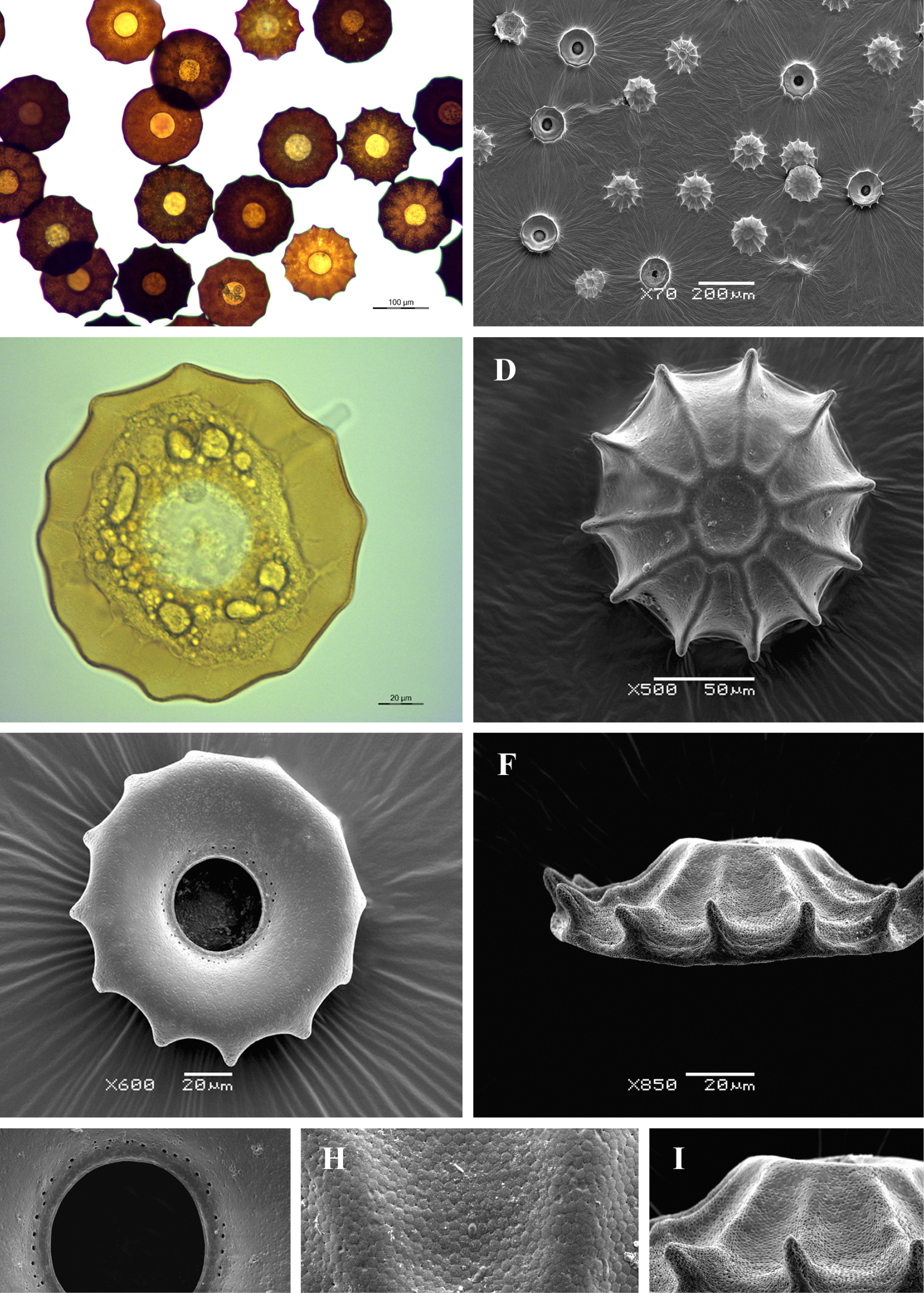
Light (A, C) and scanning electron (B, D-I) micrographs of Arcella dentata. (A, B) View of many specimens showing variability in shape and size of the shell. (C) View of live specimen illustrating two typical vesicular nuclei with one large central nucleolus, numerous small epipodes and granular endolobopodia. (D) Dodsal view showing shell depression on top and radiating ridges. (E) Apertural view. (F) Lateral view showing characteristic crown resembling shell. (G) View of aperture to illustrate small lip and surrounding large pores. (H) Portion of shell surface to show structuring alveoli and small pores. (I) Lateral view showing highly flattened shell with turned-up rim edged with conical point. |