|
||
|
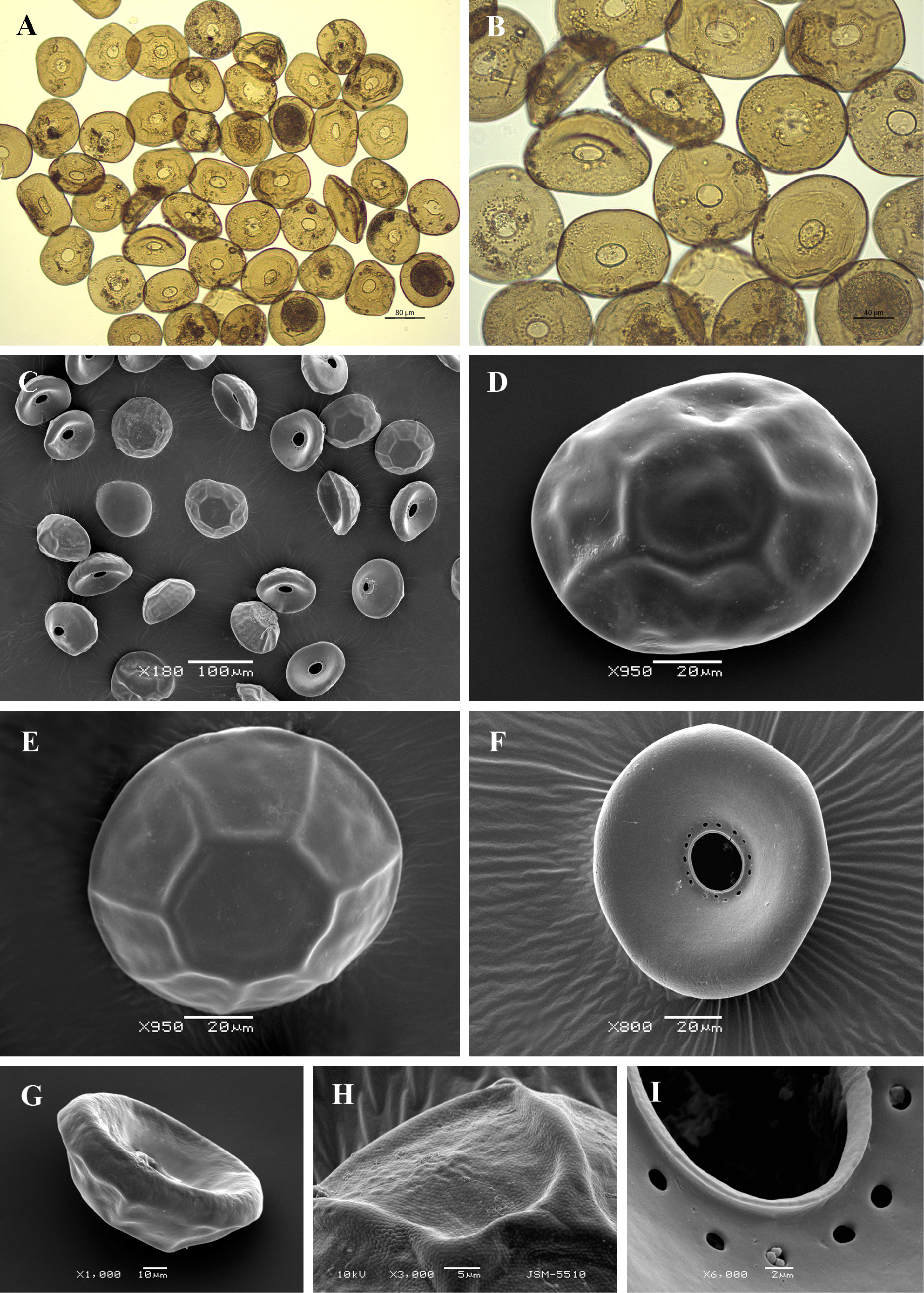
Light (A, B) and scanning electron (C-I) micrographs of Arcella catinus. (A-C) View of many specimens showing variability in shape and size of the shell. (C-E) Dorsal view to illustrate depressed aboral region and foldings of the surface; (F) Apertural view to show smooth apertural surface; (G) Lateral view showing characteristic trapezoidal shape. (H) Portion of shell surface showing small organic alveoli and foldings of the aboral surface. (I) Close up view of aperture to show distinct apertural lip and surrounding large pores. |