|
||
|
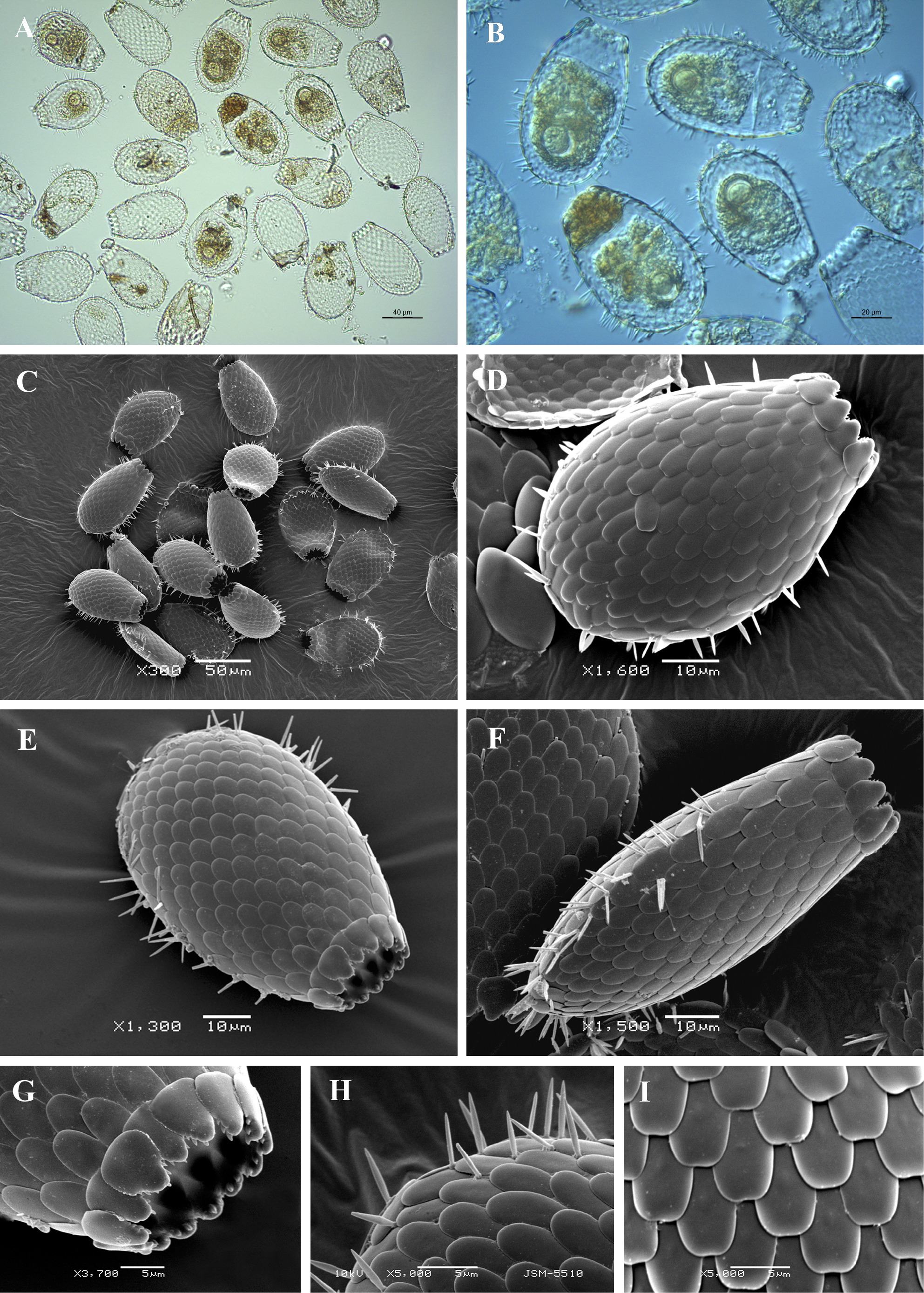
Light (A, B) and scanning electron (C-I) micrographs of Euglypha compressa. (A-C) View of many specimens to illustrate variability in shape and size of the shell. (D, E) Broad lateral view of two specimens to show general shape, arrangement of shell plates and disposition of spines. (F) Narrow lateral view. (G) Close up view of apertural region. (H) View of aboral region illustrating spines and their attachment to the shell surface. (I) Detail of shell surface to show overlapping shell-plates. |